Welcome to My blog Sites. This is your first post. Edit or delete it, then start blogging!
Month: July 2016
Week 5
This video is a good illustration of how fast bacterial cells can double:
This video shows the essential parts of a bacterial growth curve. What is the difference between the lag phase and the log phase?
[youtube https://www.youtube.com/watch?v=HP3POTboCm4]
Objective: Given a starting number of cells and a doubling time, determine the final number of cells
The formula is Final number =starting number X 2N, where N is the number of generations.
Example: I have 100 cells of a bacterial species that doubles every 30 minutes. How many cells will I have after 4 hours.
4 hours is 8 generation (30 minutes/generation with)
100 X 28=100 X 256=25,600 cells
We will do similar exercises in the labs on July 7th and 12
Objective Be able to graphically determine the doubling time of cells

This is a semi-log graph. That means that the Y axis goes up by factors of 10 with the same size intervals. In a semilog graph, the log phase of growth (when cells are doubling at constant rate) appears as a straight diagonal line). The lines between each factor increase by a unit of 1. Thus the very bottom line is 1 X 10⁷. The next line above is 2 X10⁷, the next is 3 X 10⁷ etc
Step 1: find a portion of the graph that is a straight line diagonal line. (Example the blue line)
Step 2 find 2 places on this line that differ by 2X in the Y axis. The bottom arrow is 2X 10⁸, the top arrow is 4 X 10⁸.
Step 3 determine the time for each of these 2 points. The lower point is about 2.5 hours, while the upper point is slightly less than 3 hours (say 2 hours 55 minutes). So the double time is about 25 minutes.
What is the doubling time for the red line? This will be a question on the week 5 worksheet.
Overview Video for eukaryotic cell cycle: This gives some basics on the overall process of cell reproduction and is visually striking
[youtube https://www.youtube.com/watch?v=Q6ucKWIIFmg]
Objective: Know what happens in each stage of the cell cycle including each stage of mitosis Be Able to recognize cells at each stage. We will do this in lab in week 6 but here is on line version to help you.
Be able to determine the mitotic index of cells
Be able to determine cell cycle stage lengths from data about stage distribution
Example. The total length of the cell cycle for a onion root tip cell is 30 hours. You find 120 cells in interphase, 30 in prophase, 15 in metaphase 25, in anaphase, and 15 in telophase or cytokinesis. What is the mitotic index? What is the length of prophase?
The mitotic index is the percent of total cells in mitosis. In this case, there are 200 total cells and 80 are in the stages of mitosis, so the mitotic index is 80/200=.4
To determine the length of a particular stage first determine the fraction of cells in that stage. In this case 30/200 or 15% of the cells are in prophase. We assume that the proportion of cells in a particular stage is directly related to the length of that stage (the more cells at a particular stage, the longer that stage must be. If the total cell cycle is 20 hours and 15% are in prophase, then 15% X 30=4.5 hours.
Objective: Be able to order the steps of the cell cycle and mitosis
Put The cell cycle steps in order in this image
Answers (click here)Answers
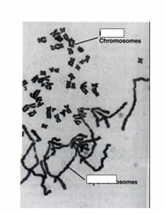
Be able to label chromosomes including sister chromatids, centromeres and telomeres.
Be able to distinguish sister chromatids from homologues

Source Own work Author Fockey003
Note that centromere, centrosome and centriole all sound alike and are easy to confuse. Only the centromere is directly associated with chromosomes. The telomere is found at the end of a chromosome and has special properties that we will talk about when we get to DNA

For a given chromosome, every individual has one homologue from each of their parents (Note that for males, the X and Y chromosome are considered homologues even though they do not have the same genes. ). A and a are different alleles (different versions of the same gene; brown and blue eyes for example)
Objective: Know the role of the following in the cell cycle: Centromere, Kinetochore, Centriole, Centrosome, Spindle, Contractile Ring
The centromere is the structure at which 2 sisters are attached. It is also attached to the Kinetochore which links the spindle to the chromosome.
This link may be of value
Centrioles are barrel shaped structures from which microtubules grow. The centrosome contains the centrioles as well as some fuzzy material called the PCM (pericentriolar material)

By Kelvinsong – Own work, CC BY 3.0, https://commons.wikimedia.org/w/index.php?curid=23107755
Plants do not typically contain centrioles, but they do contain centrosomes with the PCM. This appears sufficient to form spindles
Objective: Be able to list cell cycle checkpoints and determine the effects of defects in cell cycle checkpoints
This animation will help you understand the basic checkpoints:
Once you have done that, play this game (Click on “play the control of the cell cycle game)
Objective: Be able to predict what happens when cells from different phases are fused together. Explain the results of fusion of interphase and M cells with respect to cyclin and MPF
The key to these experiment is that something accumulates at The beginning of S phase and M phase to cause cells to go into those phases. Years of genetic and biochemical research showed that this something is a factor called cyclin. Cyclin is a protein that accumulates at particular cell cycle stages and then are destroyed. Different cyclins trigger different stages.
In this experiment
Which cyclin is important for M phase?
Cyclin by itself does not trigger M phase. Cyclin binds to a protein called CDK (Cell Division Kinase). A Kinase is an enzyme that adds phosphates to proteins. It is only active when bound to cyclin. When the appropriate cyclin binds M phase CDK, MPF (mitosis promoting factor) is formed. MPF activates mitosis by phosphorylating proteins needed for M phase events (Where might some of these proteins be found? What are their roles in mitosis?).
So what happens to cyclin and MPF when a cell in G1 is fused to a cell in M phase? How does this explain the result?
Objective: Be able to list the properties of stem cells and give some applications
Watch this video
[youtube https://www.youtube.com/watch?v=_hbgeQzmU9U]
And then go through this activity
This site shows a recent promising stem cell technique
Click on the video on the site: