Viruses

Most of this material is from Open Stax

Watch this introductory video

Amoeba Sisters Virus

Viruses are diverse entities. They vary in their structure, their replication methods, and in their target hosts. Nearly all forms of life—from bacteria and archaea to eukaryotes such as plants, animals, and fungi—have viruses that infect them. While most biological diversity can be understood through evolutionary history, such as how species have adapted to conditions and environments, much about virus origins and evolution remains unknown.

Discovery and Detection

Viruses were first discovered after the development of a porcelain filter, called the Chamberland-Pasteur filter, which could remove all bacteria visible in the microscope from any liquid sample. In 1886, Adolph Meyer demonstrated that a disease of tobacco plants, tobacco mosaic disease, could be transferred from a diseased plant to a healthy one via liquid plant extracts. In 1892, Dmitri Ivanowski showed that this disease could be transmitted in this way even after the Chamberland-Pasteur filter had removed all viable bacteria from the extract. Still, it was many years before it was proven that these “filterable” infectious agents were not simply very small bacteria but were a new type of very small, disease-causing particle.

Virions, single virus particles, are very small, about 20–250 nanometers in diameter. These individual virus particles are the infectious form of a virus outside the host cell. Unlike bacteria (which are about 100-times larger), we cannot see viruses with a light microscope, with the exception of some large virions of the poxvirus family. It was not until the development of the electron microscope in the late 1930s that scientists got their first good view of the structure of the tobacco mosaic virus (TMV) ([link]) and other viruses (Figure). The surface structure of virions can be observed by both scanning and transmission electron microscopy, whereas the internal structures of the virus can only be observed in images from a transmission electron microscope. The use of these technologies has allowed for the discovery of many viruses of all types of living organisms. They were initially grouped by shared morphology. Later, groups of viruses were classified by the type of nucleic acid they contained, DNA or RNA, and whether their nucleic acid was single- or double-stranded. More recently, molecular analysis of viral replicative cycles has further refined their classification.

Micrograph a shows a virus with a hexagonal head that stands on thin, bent legs. The virus sits on the surface of a cell that is so large that only a small fraction of its surface is visible. Micrograph b shows small bacterial cells that are about the size of the organelles in the adjacent colon cells.
In these transmission electron micrographs, (a) a virus is dwarfed by the bacterial cell it infects, while (b) these E. coli cells are dwarfed by cultured colon cells.
(credit a: modification of work by U.S. Dept. of Energy, Office of Science, LBL, PBD; credit b: modification of work by J.P. Nataro and S. Sears, unpub. data, CDC; scale-bar data from Matt Russell)

Evolution of Viruses

Although biologists have accumulated a significant amount of knowledge about how present-day viruses evolve, much less is known about how viruses originated in the first place. When exploring the evolutionary history of most organisms, scientists can look at fossil records and similar historic evidence. However, viruses do not fossilize, so researchers must conjecture by investigating how today’s viruses evolve and by using biochemical and genetic information to create speculative virus histories.

While most findings agree that viruses don’t have a single common ancestor, scholars have yet to find a single hypothesis about virus origins that is fully accepted in the field. One such hypothesis, called devolution or the regressive hypothesis, proposes to explain the origin of viruses by suggesting that viruses evolved from free-living cells. However, many components of how this process might have occurred are a mystery. A second hypothesis (called escapist or the progressive hypothesis) accounts for viruses having either an RNA or a DNA genome and suggests that viruses originated from RNA and DNA molecules that escaped from a host cell. A third hypothesis posits a system of self-replication similar to that of other self-replicating molecules, likely evolving alongside the cells they rely on as hosts; studies of some plant pathogens support this hypothesis.

As technology advances, scientists may develop and refine further hypotheses to explain the origin of viruses. The emerging field called virus molecular systematics attempts to do just that through comparisons of sequenced genetic material. These researchers hope to one day better understand the origin of viruses, a discovery that could lead to advances in the treatments for the ailments they produce.

Viral Morphology

Viruses are acellular, meaning they are biological entities that do not have a cellular structure. They therefore lack most of the components of cells, such as organelles, ribosomes, and the plasma membrane. A virion consists of a nucleic acid core, an outer protein coating or capsid, and sometimes an outer envelope made of protein and phospholipid membranes derived from the host cell. Viruses may also contain additional proteins, such as enzymes. The most obvious difference between members of viral families is their morphology, which is quite diverse. An interesting feature of viral complexity is that the complexity of the host does not correlate with the complexity of the virion. Some of the most complex virion structures are observed in bacteriophages, viruses that infect the simplest living organisms, bacteria.

Morphology

Viruses come in many shapes and sizes, but these are consistent and distinct for each viral family. All virions have a nucleic acid genome covered by a protective layer of proteins, called a capsid. The capsid is made up of protein subunits called capsomeres. Some viral capsids are simple polyhedral “spheres,” whereas others are quite complex in structure.

In general, the shapes of viruses are classified into four groups: filamentous, isometric (or icosahedral), enveloped, and head and tail. Filamentous viruses are long and cylindrical. Many plant viruses are filamentous, including TMV. Isometric viruses have shapes that are roughly spherical, such as poliovirus or herpesviruses. Enveloped viruses have membranes surrounding capsids. Animal viruses, such as HIV, are frequently enveloped. Head and tail viruses infect bacteria and have a head that is similar to icosahedral viruses and a tail shape like filamentous viruses.

Many viruses use some sort of glycoprotein to attach to their host cells via molecules on the cell called viral receptors (Figure). For these viruses, attachment is a requirement for later penetration of the cell membrane, so they can complete their replication inside the cell. The receptors that viruses use are molecules that are normally found on cell surfaces and have their own physiological functions. Viruses have simply evolved to make use of these molecules for their own replication. For example, HIV uses the CD4 molecule on T lymphocytes as one of its receptors. CD4 is a type of molecule called a cell adhesion molecule, which functions to keep different types of immune cells in close proximity to each other during the generation of a T lymphocyte immune response.

 In the illustration a viral receptor on the surface of a KSHV virus is attached to an xCT receptor embedded in the plasma membrane.
The KSHV virus binds the xCT receptor on the surface of human cells. xCT receptors protect cells against stress. Stressed cells express more xCT receptors than non-stressed cells. The KSHV virion causes cells to become stressed, thereby increasing expression of the receptor to which it binds. (credit: modification of work by NIAID, NIH)

Among the most complex virions known, the T4 bacteriophage, which infects the Escherichia coli bacterium, has a tail structure that the virus uses to attach to host cells and a head structure that houses its DNA.

Adenovirus, a non-enveloped animal virus that causes respiratory illnesses in humans, uses glycoprotein spikes protruding from its capsomeres to attach to host cells. Non-enveloped viruses also include those that cause polio (poliovirus), plantar warts (papillomavirus), and hepatitis A (hepatitis A virus).

Enveloped virions like HIV, the causative agent in AIDS, consist of nucleic acid (RNA in the case of HIV) and capsid proteins surrounded by a phospholipid bilayer envelope and its associated proteins. Glycoproteins embedded in the viral envelope are used to attach to host cells. Other envelope proteins are the matrix proteins that stabilize the envelope and often play a role in the assembly of progeny virions. Chicken pox, influenza, and mumps are examples of diseases caused by viruses with envelopes. Because of the fragility of the envelope, non-enveloped viruses are more resistant to changes in temperature, pH, and some disinfectants than enveloped viruses.

Overall, the shape of the virion and the presence or absence of an envelope tell us little about what disease the virus may cause or what species it might infect, but they are still useful means to begin viral classification (Figure).

ART CONNECTION
Illustration a shows bacteriophage T4, which houses its DNA genome in a hexagonal head. A long, straight tail extends from the bottom of the head. Tail fibers attached to the base of the tail are bent, like spider legs. In b, adenovirus houses its DNA genome in a round capsid made from many small capsomere subunits. Glycoproteins extend from the capsomere, like pins from a pincushion. In c, the HIV retrovirus houses its RNA genome and an enzyme called reverse transcriptase in a bullet-shaped capsid. A spherical viral envelope, lined with matrix proteins, surrounds the capsid. Glycoproteins extend from the viral envelope.
Viruses can be either complex in shape or relatively simple. This figure shows three relatively complex virions: the bacteriophage T4, with its DNA-containing head group and tail fibers that attach to host cells; adenovirus, which uses spikes from its capsid to bind to host cells; and HIV, which uses glycoproteins embedded in its envelope to bind to host cells. Notice that HIV has proteins called matrix proteins, internal to the envelope, which help stabilize virion shape.
(credit “bacteriophage, adenovirus”: modification of work by NCBI, NIH; credit “HIV retrovirus”: modification of work by NIAID, NIH)

Which of the following statements about virus structure is true?

  1. All viruses are encased in a viral membrane.
  2. The capsomere is made up of small protein subunits called capsids.
  3. DNA is the genetic material in all viruses.
  4. Glycoproteins help the virus attach to the host cell.

Types of Nucleic Acid

Unlike nearly all living organisms that use DNA as their genetic material, viruses may use either DNA or RNA as theirs. The virus core contains the genome or total genetic content of the virus. Viral genomes tend to be small, containing only those genes that encode proteins that the virus cannot get from the host cell. This genetic material may be single- or double-stranded. It may also be linear or circular. While most viruses contain a single nucleic acid, others have genomes that have several, which are called segments.

In DNA viruses, the viral DNA directs the host cell’s replication proteins to synthesize new copies of the viral genome and to transcribe and translate that genome into viral proteins. DNA viruses cause human diseases, such as chickenpox, hepatitis B, and some venereal diseases, like herpes and genital warts.

RNA viruses contain only RNA as their genetic material. To replicate their genomes in the host cell, the RNA viruses encode enzymes that can replicate RNA into DNA, which cannot be done by the host cell. These RNA polymerase enzymes are more likely to make copying errors than DNA polymerases, and therefore often make mistakes during transcription. For this reason, mutations in RNA viruses occur more frequently than in DNA viruses. This causes them to change and adapt more rapidly to their host. Human diseases caused by RNA viruses include hepatitis C, measles, and rabies.

Virus Classification

To understand the features shared among different groups of viruses, a classification scheme is necessary. As most viruses are not thought to have evolved from a common ancestor, however, the methods that scientists use to classify living things are not very useful. Biologists have used several classification systems in the past, based on the morphology and genetics of the different viruses. However, these earlier classification methods grouped viruses differently, based on which features of the virus they were using to classify them. The most commonly used classification method today is called the Baltimore classification scheme and is based on how messenger RNA (mRNA) is generated in each particular type of virus.

Baltimore Classification

The most commonly used system of virus classification was developed by Nobel Prize-winning biologist David Baltimore in the early 1970s. In addition to the differences in morphology and genetics mentioned above, the Baltimore classification scheme groups viruses according to how the mRNA is produced during the replicative cycle of the virus.

Group I viruses contain double-stranded DNA (dsDNA) as their genome. Their mRNA is produced by transcription in much the same way as with cellular DNA.

Group II viruses have single-stranded DNA (ssDNA) as their genome. They convert their single-stranded genomes into a dsDNA intermediate before transcription to mRNA can occur.

Group III viruses use dsRNA as their genome. The strands separate, and one of them is used as a template for the generation of mRNA using the RNA-dependent RNA polymerase encoded by the virus. Group IV viruses have ssRNA as their genome with a positive polarity. Positive polarity means that the genomic RNA can serve directly as mRNA. Intermediates of dsRNA, called replicative intermediates, are made in the process of copying the genomic RNA. Multiple, full-length RNA strands of negative polarity (complimentary to the positive-stranded genomic RNA) are formed from these intermediates, which may then serve as templates for the production of RNA with positive polarity, including both full-length genomic RNA and shorter viral mRNAs.

Coronavirus is a positive single strand RNA virus. When it infects a cell, the RNA is an mRNA which means that it can encode proteins. This RNA can be transcribed to make a negative strand RNA. The negative strand can not make proteins, but it can make more positive strands which can be put in viral particles or be used to make more protein.

Group V viruses contain ssRNA genomes with a negative polarity, meaning that their sequence is complementary to the mRNA. As with Group IV viruses, dsRNA intermediates are used to make copies of the genome and produce mRNA. In this case, the negative-stranded genome can be converted directly to mRNA. Additionally, full-length positive RNA strands are made to serve as templates for the production of the negative-stranded genome.

Group VI viruses have diploid (two copies) ssRNA genomes that must be converted, using the enzyme reverse transcriptase, to dsDNA; the dsDNA is then transported to the nucleus of the host cell and inserted into the host genome. Then, mRNA can be produced by transcription of the viral DNA that was integrated into the host genome. Group VII viruses have partial dsDNA genomes and make ssRNA intermediates that act as mRNA, but are also converted back into dsDNA genomes by reverse transcriptase, necessary for genome replication. The characteristics of each group in the Baltimore classification are summarized in Table with examples of each group.

Baltimore Classification
Group Characteristics Mode of mRNA Production Example
I Double-stranded DNA mRNA is transcribed directly from the DNA template Herpes simplex (herpesvirus)
II Single-stranded DNA DNA is converted to double-stranded form before RNA is transcribed Canine parvovirus (parvovirus)
III Double-stranded RNA mRNA is transcribed from the RNA genome Childhood gastroenteritis (rotavirus)
IV Single stranded RNA (+) Genome functions as mRNA Common cold (pircornavirus)
V Single stranded RNA (-) mRNA is transcribed from the RNA genome Rabies (rhabdovirus)
VI Single stranded RNA viruses with reverse transcriptase Reverse transcriptase makes DNA from the RNA genome; DNA is then incorporated in the host genome; mRNA is transcribed from the incorporated DNA Human immunodeficiency virus (HIV)
VII Double stranded DNA viruses with reverse transcriptase The viral genome is double-stranded DNA, but viral DNA is replicated through an RNA intermediate; the RNA may serve directly as mRNA or as a template to make mRNA


Hepatitis B virus (hepadnavirus)

  1. Viruses are tiny, acellular entities that can usually only be seen with an electron microscope. Their genomes contain either DNA or RNA—never both—and they replicate using the replication proteins of a host cell. Viruses are diverse, infecting archaea, bacteria, fungi, plants, and animals. Viruses consist of a nucleic acid core surrounded by a protein capsid with or without an outer lipid envelope. The capsid shape, presence of an envelope, and core composition dictate some elements of the classification of viruses. The most commonly used classification method, the Baltimore classification, categorizes viruses based on how they produce their mRNA.
From DNA to Protein

Intro video

What is a gene?

In both prokaryotes and eukaryotes, the second function of DNA (the first was replication) is to provide the information needed to construct the proteins necessary so that the cell can perform all of its functions. To do this, the DNA is “read” or transcribed into an mRNA molecule. The mRNA then provides the code to form a protein by a process called translation. Through the processes of transcription and translation, a protein is built with a specific sequence of amino acids that was originally encoded in the DNA. This module discusses the details of transcription.

The Central Dogma: DNA Encodes RNA; RNA Encodes Protein

The flow of genetic information in cells from DNA to mRNA to protein is described by the central dogma which states that genes specify the sequences of mRNAs, which in turn specify the sequences of proteins.

The central dogma states that DNA encodes RNA, which in turn encodes protein.

The copying of DNA to mRNA is relatively straightforward, with one nucleotide being added to the mRNA strand for every complementary nucleotide read in the DNA strand. The translation to protein is more complex because groups of three mRNA nucleotides correspond to one amino acid of the protein sequence. However, as we shall see in the next module, the translation to protein is still systematic, such that nucleotides 1 to 3 correspond to amino acid 1, nucleotides 4 to 6 correspond to amino acid 2, and so on.

RNA features

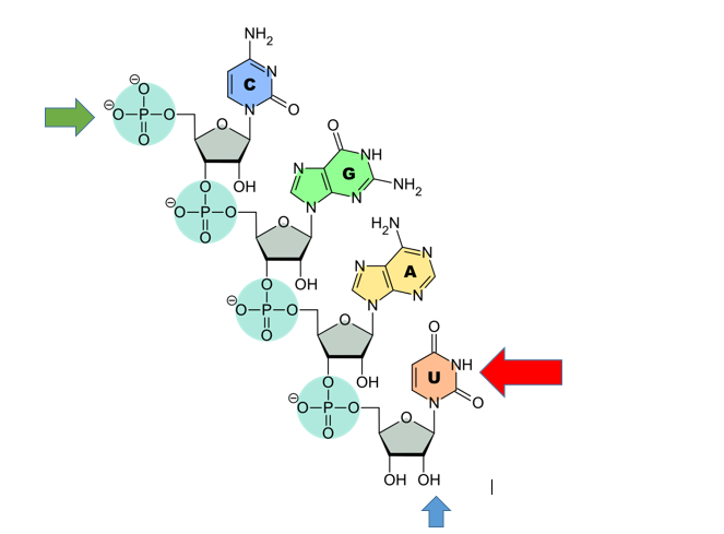
RNA contains U, not T. (Red arrow). U pairs with A

RNA contains ribose. The Blue arrow points to the ribose sugar. Note the oxygen. DNA would just have a hydrogen there. The hydroxyl group to the left is the 3’ hydroxyl (like DNA the Green arrow points to the 5’ phosphate.

Note that this RNA is single stranded. RNA usually is single stranded unlike DNA. Sometimes RNA will pair with itself.

Transcription: from DNA to mRNA

Both prokaryotes and eukaryotes perform fundamentally the same process of transcription, with the important difference of the membrane-bound nucleus in eukaryotes. With the genes bound in the nucleus, transcription occurs in the nucleus of the cell and the mRNA transcript must be transported to the cytoplasm. The prokaryotes, which include bacteria and archaea, lack membrane-bound nuclei and other organelles, and transcription occurs in the cytoplasm of the cell. In both prokaryotes and eukaryotes, transcription occurs in three main stages: initiation, elongation, and termination.

Initiation

Transcription requires the DNA double helix to partially unwind in the region of mRNA synthesis. The region of unwinding is called a transcription bubble. The DNA sequence onto which the proteins and enzymes involved in transcription bind to initiate the process is called a promoter. In most cases, promoters exist upstream of the genes they regulate. The specific sequence of a promoter is very important because it determines whether the corresponding gene is transcribed all of the time, some of the time, or hardly at all (Figure).

The initiation of transcription begins when DNA is unwound, forming a transcription bubble. Enzymes and other proteins involved in transcription bind at the promoter.

Elongation

Transcription always proceeds from one of the two DNA strands, which is called the template strand. The mRNA product is complementary to the template strand and is almost identical to the other DNA strand, called the nontemplate strand, with the exception that RNA contains a uracil (U) in place of the thymine (T) found in DNA. During elongation, an enzyme called RNA polymerase proceeds along the DNA template adding nucleotides by base pairing with the DNA template in a manner similar to DNA replication, with the difference that an RNA strand is being synthesized that does not remain bound to the DNA template. As elongation proceeds, the DNA is continuously unwound ahead of the core enzyme and rewound behind it (Figure).

During elongation, RNA polymerase tracks along the DNA template, synthesizes mRNA in the 5′ to 3′ direction, and unwinds then rewinds the DNA as it is read.

Problem example

A DNA is 3′ TAC AGC TTG 5′ What is the RNA that can be made from it?

5′ AUG UCG AAC 3′

3′ TAC AGC TTG 5′

Note that C and G pairing is just like DNA. U in RNA pairs with A in DNA. T in DNA pairs with A in RNA like DNA.

Transcription video

Transcription video


Splicing and other RNA processing

Eukaryotic RNA Processing

The newly transcribed eukaryotic mRNAs must undergo several processing steps before they can be transferred from the nucleus to the cytoplasm and translated into a protein. The additional steps involved in eukaryotic mRNA maturation create a molecule that is much more stable than a prokaryotic mRNA. For example, eukaryotic mRNAs last for several hours, whereas the typical prokaryotic mRNA lasts no more than five seconds.

The mRNA transcript is first coated in RNA-stabilizing proteins to prevent it from degrading while it is processed and exported out of the nucleus. This occurs while the pre-mRNA still is being synthesized by adding a special nucleotide “cap” to the 5′ end of the growing transcript. In addition to preventing degradation, factors involved in protein synthesis recognize the cap to help initiate translation by ribosomes.

Once elongation is complete, an enzyme then adds a string of approximately 200 adenine residues to the 3′ end, called the poly-A tail. This modification further protects the pre-mRNA from degradation and signals to cellular factors that the transcript needs to be exported to the cytoplasm.

Eukaryotic genes are composed of protein-coding sequences called exons (ex-on signifies that they are expressed) and intervening sequences called introns (int-ron denotes their intervening role). Introns are removed from the pre-mRNA during processing. Intron sequences in mRNA do not encode functional proteins. It is essential that all of a pre-mRNA’s introns be completely and precisely removed before protein synthesis so that the exons join together to code for the correct amino acids. If the process errs by even a single nucleotide, the sequence of the rejoined exons would be shifted, and the resulting protein would be nonfunctional. The process of removing introns and reconnecting exons is called splicing (Figure). Introns are removed and degraded while the pre-mRNA is still in the nucleus.

Eukaryotic mRNA contains introns that must be spliced out. A 5′ cap and 3′ tail are also added.

Note  that the RNA contained 3 exons and 2 introns.  RNAs begin and end with exons. There are always 1 more exon than intron in the primary transcript.

The video below shows the relationship between

Splicing and SMA


Why do this? What is the point of removing introns? They are not used, they are just degraded once removed. One reason is alternative splicing. IN this process, some exons cam be removed during splicing. This can yield more than one protein from the same RNA. The following lesson explains alternate splicing

Alternate Splicing


Translation

The synthesis of proteins is one of a cell’s most energy-consuming metabolic processes. In turn, proteins account for more mass than any other component of living organisms (with the exception of water), and proteins perform a wide variety of the functions of a cell. The process of translation, or protein synthesis, involves decoding an mRNA message into a polypeptide product. Amino acids are covalently strung together in lengths ranging from approximately 50 amino acids to more than 1,000.

The Protein Synthesis Machinery

In addition to the mRNA template, many other molecules contribute to the process of translation. The composition of each component may vary across species; for instance, ribosomes may consist of different numbers of ribosomal RNAs (rRNA) and polypeptides depending on the organism. However, the general structures and functions of the protein synthesis machinery are comparable from bacteria to human cells. Translation requires the input of an mRNA template, ribosomes, tRNAs, and various enzymatic factors (Figure 9.19).

Illustration of the molecules involved in protein translation. A ribosome is shown with mRNA and tRNA. Amino acids are emerging to form a protein chain.
Figure 9.19 The protein synthesis machinery includes the large and small subunits of the ribosome, mRNA, and tRNA. (credit: modification of work by NIGMS, NIH)

In E. coli, there are 200,000 ribosomes present in every cell at any given time. A ribosome is a complex macromolecule composed of structural and catalytic rRNAs, and many distinct polypeptides. In eukaryotes, the nucleolus is completely specialized for the synthesis and assembly of rRNAs.

Ribosomes are located in the cytoplasm in prokaryotes and in the cytoplasm and endoplasmic reticulum of eukaryotes. Ribosomes are made up of a large and a small subunit that come together for translation. The small subunit is responsible for binding the mRNA template, whereas the large subunit sequentially binds tRNAs, a type of RNA molecule that brings amino acids to the growing chain of the polypeptide. Each mRNA molecule is simultaneously translated by many ribosomes, all synthesizing protein in the same direction.

Depending on the species, 40 to 60 types of tRNA exist in the cytoplasm. Serving as adaptors, specific tRNAs bind to sequences on the mRNA template and add the corresponding amino acid to the polypeptide chain. Therefore, tRNAs are the molecules that actually “translate” the language of RNA into the language of proteins. For each tRNA to function, it must have its specific amino acid bonded to it. In the process of tRNA “charging,” each tRNA molecule is bonded to its correct amino acid.

The Genetic Code

To summarize what we know to this point, the cellular process of transcription generates messenger RNA (mRNA), a mobile molecular copy of one or more genes with an alphabet of A, C, G, and uracil (U). Translation of the mRNA template converts nucleotide-based genetic information into a protein product. Protein sequences consist of 20 commonly occurring amino acids; therefore, it can be said that the protein alphabet consists of 20 letters. Each amino acid is defined by a three-nucleotide sequence called the triplet codon. The relationship between a nucleotide codon and its corresponding amino acid is called the genetic code.

Given the different numbers of “letters” in the mRNA and protein “alphabets,” combinations of nucleotides corresponded to single amino acids. Using a three-nucleotide code means that there are a total of 64 (4 × 4 × 4) possible combinations; therefore, a given amino acid is encoded by more than one nucleotide triplet (Figure 9.20).

Figure shows all 64 codons. Sixty-two of these code for amino acids, and three are stop codons shown in red. The start codon, AUG, is colored green.
Figure 9.20This figure shows the genetic code for translating each nucleotide triplet, or codon, in mRNA into an amino acid or a termination signal in a nascent protein. (credit: modification of work by NIH)

Three of the 64 codons terminate protein synthesis and release the polypeptide from the translation machinery. These triplets are called stop codons. Another codon, AUG, also has a special function. In addition to specifying the amino acid methionine, it also serves as the start codon to initiate translation. The reading frame for translation is set by the AUG start codon near the 5′ end of the mRNA. The genetic code is universal. With a few exceptions, virtually all species use the same genetic code for protein synthesis, which is powerful evidence that all life on Earth shares a common origin.

Problem

Problem example

A DNA is 3′ TAC AGC TTG 5′ What is the Protein that can be made from it?

5′ AUG UCG AAC 3′ is the RNA

MET  CYS  ASN is the amino acid sequence

You will not have to memorize the codon table

You will have to be able to use it


The Mechanism of Protein Synthesis

Just as with mRNA synthesis, protein synthesis can be divided into three phases: initiation, elongation, and termination. The process of translation is similar in prokaryotes and eukaryotes. Here we will explore how translation occurs in E. coli, a representative prokaryote, and specify any differences between prokaryotic and eukaryotic translation.

Protein synthesis begins with the formation of an initiation complex. In E. coli, this complex involves the small ribosome subunit, the mRNA template, three initiation factors, and a special initiator tRNA. The initiator tRNA interacts with the AUG start codon, and links to a special form of the amino acid methionine that is typically removed from the polypeptide after translation is complete.

In prokaryotes and eukaryotes, the basics of polypeptide elongation are the same, so we will review elongation from the perspective of E. coli. The large ribosomal subunit of E. coli consists of three compartments: the A site binds incoming charged tRNAs (tRNAs with their attached specific amino acids). The P site binds charged tRNAs carrying amino acids that have formed bonds with the growing polypeptide chain but have not yet dissociated from their corresponding tRNA. The E site releases dissociated tRNAs so they can be recharged with free amino acids. The ribosome shifts one codon at a time, catalyzing each process that occurs in the three sites. With each step, a charged tRNA enters the complex, the polypeptide becomes one amino acid longer, and an uncharged tRNA departs. The energy for each bond between amino acids is derived from GTP, a molecule similar to ATP (Figure 9.21). Amazingly, the E. coli translation apparatus takes only 0.05 seconds to add each amino acid, meaning that a 200-amino acid polypeptide could be translated in just 10 seconds.

Illustration shows the steps of protein synthesis. First, an initiator tRNA recognizes the sequence AUG on the mRNA that is associated with the small ribosomal subunit. The large subunit joins the complex. Next, a second tRNA is recruited at the A site. A peptide bond is formed between the first amino acid, which is at the P site, and the second amino acid, which is at the A site. The mRNA then shifts and the first tRNA is moved to the E site, where it dissociates from the ribosome. Another tRNA binds the A site, and the process is repeated.
Figure 9.21 Translation begins when a tRNA anticodon recognizes a codon on the mRNA. The large ribosomal subunit joins the small subunit, and a second tRNA is recruited. As the mRNA moves relative to the ribosome, the polypeptide chain is formed. Entry of a release factor into the A site terminates translation and the components dissociate.


DNA

Much of this material is from Open Stax

Intro video

Evidence that DNA is the genetic material 

Our current understanding of DNA began with the discovery of nucleic acids followed by the development of the double-helix model. In the 1860s, Friedrich Miescher (Figure 14.2), a physician by profession, isolated phosphate-rich chemicals from white blood cells (leukocytes). He named these chemicals (which would eventually be known as DNA) nuclein because they were isolated from the nuclei of the cells.

Photo of Friedrich Miescher.
Figure 14.2 Friedrich Miescher (1844–1895) discovered nucleic acids.

To see Miescher conduct his experiment that led to his discovery of DNA and associated proteins in the nucleus, click through this review.

A half century later, in 1928, British bacteriologist Frederick Griffith reported the first demonstration of bacterial transformation—a process in which external DNA is taken up by a cell, thereby changing its morphology and physiology. Griffith conducted his experiments with Streptococcus pneumoniae, a bacterium that causes pneumonia. Griffith worked with two strains of this bacterium called rough (R) and smooth (S). (The two cell types were called “rough” and “smooth” after the appearance of their colonies grown on a nutrient agar plate.)

The R strain is non-pathogenic (does not cause disease). The S strain is pathogenic (disease-causing), and has a capsule outside its cell wall. The capsule allows the cell to escape the immune responses of the host mouse.

When Griffith injected the living S strain into mice, they died from pneumonia. In contrast, when Griffith injected the live R strain into mice, they survived. In another experiment, when he injected mice with the heat-killed S strain, they also survived. This experiment showed that the capsule alone was not the cause of death. In a third set of experiments, a mixture of live R strain and heat-killed S strain were injected into mice, and—to his surprise—the mice died. Upon isolating the live bacteria from the dead mouse, only the S strain of bacteria was recovered. When this isolated S strain was injected into fresh mice, the mice died. Griffith concluded that something had passed from the heat-killed S strain into the live R strain and transformed it into the pathogenic S strain. He called this the transforming principle (Figure 14.3). These experiments are now known as Griffith’s transformation experiments.

On the left is a photo of a live mouse, representing a mouse injected with heat-killed, virulent S strain. On the right is a photo of a dead mouse, representing a mouse injected with heat-killed, virulent S strain and live, non-virulent R strain.
Figure 14.3 Two strains of S. pneumoniae were used in Griffith’s transformation experiments. The R strain is non-pathogenic, whereas the S strain is pathogenic and causes death. When Griffith injected a mouse with the heat-killed S strain and a live R strain, the mouse died. The S strain was recovered from the dead mouse. Griffith concluded that something had passed from the heat-killed S strain to the R strain, transforming the R strain into the S strain in the process. (credit “living mouse”: modification of work by NIH; credit “dead mouse”: modification of work by Sarah Marriage)

Scientists Oswald Avery, Colin MacLeod, and Maclyn McCarty (1944) were interested in exploring this transforming principle further. They isolated the S strain from the dead mice and isolated the proteins and nucleic acids (RNA and DNA) as these were possible candidates for the molecule of heredity. They used enzymes that specifically degraded each component and then used each mixture separately to transform the R strain. They found that when DNA was degraded, the resulting mixture was no longer able to transform the bacteria, whereas all of the other combinations were able to transform the bacteria. This led them to conclude that DNA was the transforming principle.

A second set of experiments showed that this is a more general principle. The experiment linked here shows that the genetic material in a bacterial virus is DNA. For this activity:

Click on the link

Click on “Animation”

Click on “jump to” (lower left)

Click on the second circle from the right

The Hershey Chase Experiment

Around this same time, Austrian biochemist Erwin Chargaff examined the content of DNA in different species and found that the amounts of adenine, thymine, guanine, and cytosine were not found in equal quantities, and that relative concentrations of the four nucleotide bases varied from species to species, but not within tissues of the same individual or between individuals of the same species. He also discovered something unexpected: That the amount of adenine equaled the amount of thymine, and the amount of cytosine equaled the amount of guanine (that is, A = T and G = C). Different species had equal amounts of purines (A+G) and pyrimidines (T + C), but different ratios of A+T to G+C. These observations became known as Chargaff’s rules. Chargaff’s findings proved immensely useful when Watson and Crick were getting ready to propose their DNA double helix model! You can see after reading the past few pages how science builds upon previous discoveries, sometimes in a slow and laborious process.

Sample Problem
An organism has 22% A. What percent T, C, and G does it have?
(1)A=T =22%
(2) A+T+C+G=100%
(3) 22+22 +C +G =100%
(4) C+G= 100-44= 56%
(5) C=G= 28%
DNA Structure

The building blocks of DNA are nucleotides. The important components of the nucleotide are a nitrogenous (nitrogen-bearing) base, a 5-carbon sugar (pentose), and a phosphate group (Figure 14.5). The nucleotide is named depending on the nitrogenous base. The nitrogenous base can be a purine such as adenine (A) and guanine (G), or a pyrimidine such as cytosine (C) and thymine (T).

VISUAL CONNECTION

Illustration depicts the structure of a nucleoside, which is made up of a pentose with a nitrogenous base attached at the 1 prime position. There are two kinds of nitrogenous bases: pyrimidines, which have one six-membered ring, and purines, which have a six-membered ring fused to a five-membered ring. Cytosine, thymine, and uracil are pyrimidines, and adenine and guanine are purines. A nucleoside with a phosphate attached at the 5 prime position is called a mononucleotide. A nucleoside with two or three phosphates attached is called a nucleotide diphosphate or nucleotide triphosphate, respectively.
Figure 14.5 The purines have a double ring structure with a six-membered ring fused to a five-membered ring. Pyrimidines are smaller in size; they have a single six-membered ring structure.

The images above illustrate the five bases of DNA and RNA. Examine the images and explain why these are called “nitrogenous bases.” How are the purines different from the pyrimidines? How is one purine or pyrimidine different from another, e.g., adenine from guanine? How is a nucleoside different from a nucleotide?

The purines have a double ring structure with a six-membered ring fused to a five-membered ring. Pyrimidines are smaller in size; they have a single six-membered ring structure.

The sugar is deoxyribose in DNA and ribose in RNA. The carbon atoms of the five-carbon sugar are numbered 1′, 2′, 3′, 4′, and 5′ (1′ is read as “one prime”). The phosphate, which makes DNA and RNA acidic, is connected to the 5′ carbon of the sugar by the formation of an ester linkage between phosphoric acid and the 5′-OH group (an ester is an acid + an alcohol). In DNA nucleotides, the 3′ carbon of the sugar deoxyribose is attached to a hydroxyl (OH) group. In RNA nucleotides, the 2′ carbon of the sugar ribose also contains a hydroxyl group. The base is attached to the 1’carbon of the sugar.

The nucleotides combine with each other to produce phosphodiester bonds. The phosphate residue attached to the 5′ carbon of the sugar of one nucleotide forms a second ester linkage with the hydroxyl group of the 3′ carbon of the sugar of the next nucleotide, thereby forming a 5′-3′ phosphodiester bond. In a polynucleotide, one end of the chain has a free 5′ phosphate, and the other end has a free 3′-OH. These are called the 5′ and 3′ ends of the chain.

In the 1950s, Francis Crick and James Watson worked together to determine the structure of DNA at the University of Cambridge, England. Other scientists like Linus Pauling and Maurice Wilkins were also actively exploring this field. Pauling previously had discovered the secondary structure of proteins using X-ray crystallography. In Wilkins’ lab, researcher Rosalind Franklin was using X-ray diffraction methods to understand the structure of DNA. Watson and Crick were able to piece together the puzzle of the DNA molecule on the basis of Franklin’s data because Crick had also studied X-ray diffraction (Figure 14.6). In 1962, James Watson, Francis Crick, and Maurice Wilkins were awarded the Nobel Prize in Medicine. Unfortunately, by then Franklin had died, and Nobel prizes are not awarded posthumously.

The photo in part A shows James Watson, Francis Crick, and Maclyn McCarty. The x-ray diffraction pattern in part b is symmetrical, with dots in an x-shape
Figure 14.6 The work of pioneering scientists (a) James Watson, Francis Crick, and Maclyn McCarty led to our present day understanding of DNA. Scientist Rosalind Franklin discovered (b) the X-ray diffraction pattern of DNA, which helped to elucidate its double-helix structure. (credit a: modification of work by Marjorie McCarty, Public Library of Science)

Watson and Crick proposed that DNA is made up of two strands that are twisted around each other to form a right-handed helix. Base pairing takes place between a purine and pyrimidine on opposite strands, so that A pairs with T, and G pairs with C (suggested by Chargaff’s Rules). Thus, adenine and thymine are complementary base pairs, and cytosine and guanine are also complementary base pairs. The base pairs are stabilized by hydrogen bonds: adenine and thymine form two hydrogen bonds and cytosine and guanine form three hydrogen bonds. The two strands are anti-parallel in nature; that is, the 3′ end of one strand faces the 5′ end of the other strand. The sugar and phosphate of the nucleotides form the backbone of the structure, whereas the nitrogenous bases are stacked inside, like the rungs of a ladder. Each base pair is separated from the next base pair by a distance of 0.34 nm, and each turn of the helix measures 3.4 nm. Therefore, 10 base pairs are present per turn of the helix. The diameter of the DNA double-helix is 2 nm, and it is uniform throughout. Only the pairing between a purine and pyrimidine and the antiparallel orientation of the two DNA strands can explain the uniform diameter. The twisting of the two strands around each other results in the formation of uniformly spaced major and minor grooves (Figure 14.7).

Part A shows an illustration of a D N A double helix, which has a sugar phosphate backbone on the outside and nitrogenous base pairs on the inside. Part B shows base pairing between thymine and adenine, which form two hydrogen bonds, and between guanine and cytosine, which form three hydrogen bonds. Part C shows a molecular model of the D N A double helix. The outside of the helix alternates between wide gaps, called major grooves, and narrow gaps, called minor grooves.
Figure 14.7 DNA has (a) a double helix structure and (b) phosphodiester bonds; the dotted lines between Thymine and Adenine and Guanine and Cytosine represent hydrogen bonds. The (c) major and minor grooves are binding sites for DNA binding proteins during processes such as transcription (the copying of RNA from DNA) and replication.


When a cell divides, it is important that each daughter cell receives an identical copy of the DNA. This is accomplished by the process of DNA replication. The replication of DNA occurs during the synthesis phase, or S phase, of the cell cycle, before the cell enters mitosis or meiosis.

The elucidation of the structure of the double helix provided a hint as to how DNA is copied. Recall that adenine nucleotides pair with thymine nucleotides, and cytosine with guanine. This means that the two strands are complementary to each other. For example, a strand of DNA with a nucleotide sequence of AGTCATGA will have a complementary strand with the sequence TCAGTACT

The two strands of DNA are complementary, meaning the sequence of bases in one strand can be used to create the correct sequence of bases in the other strand.


DNA Replication

The elucidation of the structure of the double helix provided a hint as to how DNA divides and makes copies of itself. In their 1953 paper, Watson and Crick penned an incredible understatement: “It has not escaped our notice that the specific pairing we have postulated immediately suggests a possible copying mechanism for the genetic material.” With specific base pairs, the sequence of one DNA strand can be predicted from its complement. The double-helix model suggests that the two strands of the double helix separate during replication, and each strand serves as a template from which the new complementary strand is copied. What was not clear was how the replication took place. There were three models suggested (Figure 14.12): conservative, semi-conservative, and dispersive.

Illustration shows the conservative, semi-conservative, and dispersive models of D N A synthesis. In the conservative model, the D N A is replicated and both newly synthesized strands are paired together. In the semi-conservative model, each newly synthesized strand pairs with a parent strand. In the dispersive model, newly synthesized D N A is interspersed with parent D N A within both D N A strands.
Figure 14.12 The three suggested models of DNA replication. Gray indicates the original DNA strands, and blue indicates newly synthesized DNA.

In conservative replication, the parental DNA remains together, and the newly formed daughter strands are together. The semi-conservative method suggests that each of the two parental DNA strands acts as a template for new DNA to be synthesized; after replication, each double-stranded DNA includes one parental or “old” strand and one “new” strand. In the dispersive model, both copies of DNA have double-stranded segments of parental DNA and newly synthesized DNA interspersed.

Later experiments showed that the semi-conservative model (suggested but not proved by Watson and Crick) is correct.

During DNA replication, each strand serves as a template from which new strands are copied. The new strands will be complementary to the parental or “old” strands. When two daughter DNA copies are formed, they have the same sequence and are divided equally into the two daughter cells.

Watch this video to understand DNA replication

DNA Replication Video

DNA Replication in Eukaryotes

Because eukaryotic genomes are very complex, DNA replication is a very complicated process that involves several enzymes and other proteins. It occurs in three main stages: initiation, elongation, and termination.

Recall that eukaryotic DNA is bound to proteins known as histones to form structures called nucleosomes. During initiation, the DNA is made accessible to the proteins and enzymes involved in the replication process. How does the replication machinery know where on the DNA double helix to begin? It turns out that there are specific nucleotide sequences called origins of replication at which replication begins. Certain proteins bind to the origin of replication while an enzyme called helicase unwinds and opens up the DNA helix. As the DNA opens up, Y-shaped structures called replication forks are formed (Figure). Two replication forks are formed at the origin of replication, and these get extended in both directions as replication proceeds. There are multiple origins of replication on the eukaryotic chromosome, such that replication can occur simultaneously from several places in the genome.

During elongation, an enzyme called DNA polymerase adds DNA nucleotides to the 3′ end of the template. Because DNA polymerase can only add new nucleotides at the end of a backbone, a primer sequence, which provides this starting point, is added with complementary RNA nucleotides. This primer is removed later, and the nucleotides are replaced with DNA nucleotides. One strand, which is complementary to the parental DNA strand, is synthesized continuously toward the replication fork so the polymerase can add nucleotides in this direction. This continuously synthesized strand is known as the leading strand. Because DNA polymerase can only synthesize DNA in a 5′ to 3′ direction, the other new strand is put together in short pieces called Okazaki fragments. The Okazaki fragments each require a primer made of RNA to start the synthesis. The strand with the Okazaki fragments is known as the lagging strand. As synthesis proceeds, an enzyme removes the RNA primer, which is then replaced with DNA nucleotides, and the gaps between fragments are sealed by an enzyme called DNA ligase.

The process of DNA replication can be summarized as follows:

  1. DNA unwinds at the origin of replication.
  2. New bases are added to the complementary parental strands. One new strand is made continuously, while the other strand is made in pieces.
  3. Primers are removed, new DNA nucleotides are put in place of the primers and the backbone is sealed by DNA ligase.


A replication fork is formed by the opening of the origin of replication, and helicase separates the DNA strands. An RNA primer is synthesized, and is elongated by the DNA polymerase. On the leading strand, DNA is synthesized continuously, whereas on the lagging strand, DNA is synthesized in short stretches. The DNA fragments are joined by DNA ligase

Here is a second DNA video:

DNA replication Detail

Meiosis 2020

Meiosis is a tricky concept. To help to you understand it, go through the following activities in order

Start by watching this video (You will use this to do this week’s class lesson)
Meiosis Video

Here are the objectives for this unit. lets take them one at a time

Be able to distinguish Diploid and haploid cells

Most of the cells in our body are diploid, they have 2 copies of each chromosome. Our diploid number is 46. Sperm and egg are haploid cells,they only have 1 copy. At the end of meiosis 1, each cell is also haploid. For example, in females, after meiosis 1 there is only a single X chromosome. Note that it has 2 sisters which will be attached until meiosis 2. In males, there will be an X or a Y (but not both) after meiosis 1. Again this chromosome will have 2 sisters.

2. Be able to determine whether a cell is in Mitosis, Meiosis 1 or Meiosis 2

To be able to do this, one first needs to be able to distinguish sisters and homologues as shown here

Sisters and Homologs in Mitosis


The Sisters are genetically identical (before recombination, which we will ignore for the moment)

The homologues contain the same genes but can contain different genetic information (for example A versus B blood type)

In M1, the homologues consist of 2 sisters which remain attached to each other. The homologues separate from each other in M1. Thus all M1 cells will show a row of 4 sisters as shown in the bottom of the figure. You will not see this in any other stage. Thus it is easy to distinguish M1 from other stages.

In M2, the sisters separate. The homologues have already separated. Thus one should never see both homolgoues in an M2 cell

Now how do we distinguish M1 from mitosis

(1) Mitotic cells contain all chromosomes that an individual has. Therefore

(A) For each chromosome they should show one homologue from their mother and one from their father. Meiotic cells show only the maternal or paternal chromosome, not both

(B)   Because of (A) mitotic cells will always have an even number of chromosomes. Cells in M2 can have an odd number of chromosomes. During anaphase, a mitotic cell will have an even number of chromosomes on each side of the metaphase plate, an M2 cells can have an odd number on each side of the plate. (this is true for an organism such as humans for which the total number of chromosomes is an odd number X 2. (46 for humans).

(C) Recombination does not occur in mitosis. If you see a recombinant chromosome (partly one color and partly another), it must not be in mitosis.

(D) If X and Y chromosomes are present (This will be indicated) A mitotic cell can have both the X and the Y (if it is male) but a cell in M2 can only have the X or the Y.

Here is some practice for you

Meiosis Test Quiz

3. Be able to compare mitosis and meiosis in terms of processes and end results

The purpose of mitosis is to create cells that are genetically identical to the parent cells. Mitosis gives you cells that have 2 copies of each chromosome, just like the parent cells

The purpose of meiosis is to create cells that have 1 (not 2) copies of each chromosome so that after fertilization, the embryo will have 2 copies of each chromosome.

In both process, there is a preceding interphase which includes chromosome duplication.

Both processes have prophase, metaphase Anaphase and Telophase using the same cellular structures such as spindles and centrioles. Meiosis has 2 sets of these stages.

In order to assure that egg and sperm only get one of each chromosome, the homologues (members of a pair) line up during M1 and separate. During M1, crossing over occurs between homologues, this results in increased genetic variation

Crossing over does not occur in mitosis.


Chromosome Duplication (Before meiosis) Then Pairing of Chromosomes (Prophase of M1) Crossing Over (Prophase of M1) separation of Homologues (Anaphase of M1)   separation of sisters (Anaphase of M2)

Think you know it? Try this?

https://h5p.org/node/148060

Know how meiosis leads to genetic variation (Know how recombination and independent assortment can increase the variation in gametes

Independent Assortment


Independent Assortment:  4 different combinations can result from 2 pairs of chromosomes.  In general the number of combinations is 2N where N is the number of pairs of chromosomes


css.php