Be able to determine whether a compound is oxidized or reduced in an oxidation reduction reaction.
In some reactions, electrons are transferred from one atom or molecule to another or they get closer to one atom than another (they form polar bonds). The atom (or molecule) receiving the electron is said to get reduced (remember electrons have a negative charge so gaining electrons makes their overall charge more negative). The atom (or molecule) losing the electrons is said to be oxidized. The following can help you remember
Oxidation is loss of electrons, Reduction is Gain (OIL RIG)
2K + Cl2→2KCl in KCl, K has a positive charge (K+) thus is oxidized relative to K,
In KCl, Cl has a negative charge (Cl–) thus is reduced relative to to Cl2.
Some more rules:
Losing Hydrogen =oxidation
Gaining Hydrogen =reduction
Losing oxygen=reduction
Gaining oxygen=oxidation
Example
6CO2 + 6H2O→C6H12O6 + 6O2 (Equation for photosynthesis)
Carbon dioxide gains hydrogen and is reduced
Water loses hydrogens and is oxidized
Try the following (more on the group worksheet)
In the following reactions which compound or element is oxidized and which is reduced?
2Ca + O2 →2CaO
CH3CH2OH + 3O2 → 2CO2 + 3H2O
For the following know (1) Where they occur (2) what is produced and what is used (ATP, NADH, carbon dioxide etc)—Glycolysis, Krebs Cycle(including the preparation step) , Oxidative Phosphorylation
Know the order of events in respiration
In the activity below you will see terms as well as steps. Drag “step 1” to the process which is the first step among those listed. If you are correct, the step and process will disappear. Continue until all of the steps are gone. How fast can you do this?
Be able to rank the energy levels for different components.
Remember the rules of thermodynamics. If 1 molecule of compound A is used to make 10 molecules of compound B, then (if there are no other energy sources) compound A must have more energy per molecule than compound B. Use this worksheet to help you:
For each of the following, know where they are made and where they use (The light cycle or Calvin Cycle): ATP, NADPH, NADP+, Oxygen, Carbon Dioxide, Sugars.
Be able to diagram a chloroplast and show where the Calvin cycles and light cycles take place.
Use this link to diagram and a label chloroplast and a mitochondrion and fill in the table in the second slide. Each of these 3 can be done for worksheet credit. To draw freehand use the “scribble” function which can be found using the tiny arrow next to the diagonal line under “tools”. This exercise is similar to the cell drawing exercise we did in class. The site we used can not be use for this exercise since it goes away when closed.
This video is a good illustration of how fast bacterial cells can double:
This video shows the essential parts of a bacterial growth curve. What is the difference between the lag phase and the log phase?
[youtube https://www.youtube.com/watch?v=HP3POTboCm4]
Objective: Given a starting number of cells and a doubling time, determine the final number of cells
The formula is Final number =starting number X 2N, where N is the number of generations.
Example: I have 100 cells of a bacterial species that doubles every 30 minutes. How many cells will I have after 4 hours.
4 hours is 8 generation (30 minutes/generation with)
100 X 28=100 X 256=25,600 cells
We will do similar exercises in the labs on July 7th and 12
Objective Be able to graphically determine the doubling time of cells
This is a semi-log graph. That means that the Y axis goes up by factors of 10 with the same size intervals. In a semilog graph, the log phase of growth (when cells are doubling at constant rate) appears as a straight diagonal line). The lines between each factor increase by a unit of 1. Thus the very bottom line is 1 X 10⁷. The next line above is 2 X10⁷, the next is 3 X 10⁷ etc
Step 1: find a portion of the graph that is a straight line diagonal line. (Example the blue line)
Step 2 find 2 places on this line that differ by 2X in the Y axis. The bottom arrow is 2X 10⁸, the top arrow is 4 X 10⁸.
Step 3 determine the time for each of these 2 points. The lower point is about 2.5 hours, while the upper point is slightly less than 3 hours (say 2 hours 55 minutes). So the double time is about 25 minutes.
What is the doubling time for the red line? This will be a question on the week 5 worksheet.
Overview Video for eukaryotic cell cycle: This gives some basics on the overall process of cell reproduction and is visually striking
[youtube https://www.youtube.com/watch?v=Q6ucKWIIFmg]
Objective: Know what happens in each stage of the cell cycle including each stage of mitosis Be Able to recognize cells at each stage. We will do this in lab in week 6 but here is on line version to help you.
Be able to determine cell cycle stage lengths from data about stage distribution
Example. The total length of the cell cycle for a onion root tip cell is 30 hours. You find 120 cells in interphase, 30 in prophase, 15 in metaphase 25, in anaphase, and 15 in telophase or cytokinesis. What is the mitotic index? What is the length of prophase?
The mitotic index is the percent of total cells in mitosis. In this case, there are 200 total cells and 80 are in the stages of mitosis, so the mitotic index is 80/200=.4
To determine the length of a particular stage first determine the fraction of cells in that stage. In this case 30/200 or 15% of the cells are in prophase. We assume that the proportion of cells in a particular stage is directly related to the length of that stage (the more cells at a particular stage, the longer that stage must be. If the total cell cycle is 20 hours and 15% are in prophase, then 15% X 30=4.5 hours.
Objective: Be able to order the steps of the cell cycle and mitosis
Be able to label chromosomes including sister chromatids, centromeres and telomeres.
Be able to distinguish sister chromatids from homologues
Source Own work Author Fockey003
Note that centromere, centrosome and centriole all sound alike and are easy to confuse. Only the centromere is directly associated with chromosomes. The telomere is found at the end of a chromosome and has special properties that we will talk about when we get to DNA
For a given chromosome, every individual has one homologue from each of their parents (Note that for males, the X and Y chromosome are considered homologues even though they do not have the same genes. ). A and a are different alleles (different versions of the same gene; brown and blue eyes for example)
Objective: Know the role of the following in the cell cycle: Centromere, Kinetochore, Centriole, Centrosome, Spindle, Contractile Ring
The centromere is the structure at which 2 sisters are attached. It is also attached to the Kinetochore which links the spindle to the chromosome.
Centrioles are barrel shaped structures from which microtubules grow. The centrosome contains the centrioles as well as some fuzzy material called the PCM (pericentriolar material)
By Kelvinsong – Own work, CC BY 3.0, https://commons.wikimedia.org/w/index.php?curid=23107755
Plants do not typically contain centrioles, but they do contain centrosomes with the PCM. This appears sufficient to form spindles
Objective: Be able to list cell cycle checkpoints and determine the effects of defects in cell cycle checkpoints
This animation will help you understand the basic checkpoints:
Objective: Be able to predict what happens when cells from different phases are fused together. Explain the results of fusion of interphase and M cells with respect to cyclin and MPF
The key to these experiment is that something accumulates at The beginning of S phase and M phase to cause cells to go into those phases. Years of genetic and biochemical research showed that this something is a factor called cyclin. Cyclin is a protein that accumulates at particular cell cycle stages and then are destroyed. Different cyclins trigger different stages.
Cyclin by itself does not trigger M phase. Cyclin binds to a protein called CDK (Cell Division Kinase). A Kinase is an enzyme that adds phosphates to proteins. It is only active when bound to cyclin. When the appropriate cyclin binds M phase CDK, MPF (mitosis promoting factor) is formed. MPF activates mitosis by phosphorylating proteins needed for M phase events (Where might some of these proteins be found? What are their roles in mitosis?).
So what happens to cyclin and MPF when a cell in G1 is fused to a cell in M phase? How does this explain the result?
Objective: Be able to list the properties of stem cells and give some applications
Watch this video
[youtube https://www.youtube.com/watch?v=_hbgeQzmU9U]
Be able to determine whether a compound is oxidized or reduced in an oxidation reduction reaction.
In some reactions, electrons are transferred from one atom or molecule to another or they get closer to one atom than another (they form polar bonds). The atom (or molecule) receiving the electron is said to get reduced (remember electrons have a negative charge so gaining electrons makes their overall charge more negative). The atom (or molecule) losing the electrons is said to be oxidized. The following can help you remember
Oxidation is loss of electrons, Reduction is Gain (OIL RIG)
2K + Cl2→2KCl in KCl, K has a positive charge (K+) thus is oxidized relative to K,
In KCl, Cl has a negative charge (Cl–) thus is reduced relative to to Cl2.
Some more rules:
Losing Hydrogen =oxidation
Gaining Hydrogen =reduction
Losing oxygen=reduction
Gaining oxygen=oxidation
Example
6CO2 + 6H2O→C6H12O6 + 6O2 (Equation for photosynthesis)
Carbon dioxide gains hydrogen and is reduced
Water loses hydrogens and is oxidized
Try the following (more on the group worksheet)
In the following reactions which compound or element is oxidized and which is reduced?
2Ca + O2 →2CaO
CH3CH2OH + 3O2 → 2CO2 + 3H2O
For the following know (1) Where they occur (2) what is produced and what is used (ATP, NADH, carbon dioxide etc)—Glycolysis, Krebs Cycle(including the preparation step) , Oxidative Phosphorylation
Here are some useful exercises on Glycolysis the Krebs Cycle and Oxidative Phosphorylation
In the activity below you will see terms as well as steps. Drag “step 1” to the process which is the first step among those listed. If you are correct, the step and process will disappear. Continue until all of the steps are gone. How fast can you do this?
Be able to rank the energy levels for different components.
Remember the rules of thermodynamics. If 1 molecule of compound A is used to make 10 molecules of compound B, then (if there are no other energy sources) compound A must have more energy per molecule than compound B. Use this worksheet to help you:
For each of the following, know where they are made and where they use (The light cycle or Calvin Cycle): ATP, NADPH, NADP+, Oxygen, Carbon Dioxide, Sugars.
Be able to draw out and analyze experiments as shown on the photosynthesis worksheet. See the group worksheet. You also look at this experiment which is relevant to both chapters 9 and 10
Be able to interpret an action spectrum and distinguish it from an absorbance spectrum
Plants move toward light. The image below shows how the relationship between the wavelength of light and the degree of movement. This is an action spectrum, because it measures a plant response as a function of wavelength. This can be compared to an absorbance spectrum which measures absorbance of light as a function of wavelength (like our first lab). What would you predict about the absorbance spectrum of a pigment that was responsible for the response in the graph below? What would be a good title for this graph. (Answer these questions and submit as a reply to this blog for worksheet credit)
Be able to diagram a chloroplast and show where the Calvin cycles and light cycles take place.
Use this link to diagram and a label chloroplast and a mitochondrion and fill in the table in the second slide. Each of these 3 can be done for worksheet credit. To draw freehand use the “scribble” function which can be found using the tiny arrow next to the diagonal line under “tools”. This exercise is similar to the cell drawing exercise we did in class. The site we used can not be use for this exercise since it goes away when closed.
Know what substrates are and how they relate to enzyme catalyzed reactions.
Example what is the substrate in this enzyme catalyzed reaction described in the video?
The substrate is the reactant (or reactants) in a reaction involving an enzyme. The reactant fits into the active site of an enzyme which in this reaction is trypsin. Note that the enzyme is shown above the arrow. Doing this helps remind you that the enzyme is not changed by the reaction. One enzyme can do many reactions (like a machine)
Know how enzymes catalyze biological reactions, specifically their effects on activation energy and their mechanism of action.
This shows how enzymes speed up reactions: They can get molecules closer to each other and in the right orientation.
Another concept in how enzymes speed up reactions is activation energy. Imagine you have a homework assignment due next week. You could do it now, or you could put it off until right before it is due. The activation energy is the energy needed to get the process started. Note that the homework might take you an hour whether you do it now or later. The activation energy is just what it takes to get started. Enzymes decrease activation energy as shown in the diagram below.
By Originally uploaded by Jerry Crimson Mann, vectorized by Tutmosis, corrected by Fvasconcellos – en:Image:Activation2.png, CC BY-SA 3.0, https://commons.wikimedia.org/w/index.php?curid=1407225
Note that that the ΔG (The overall energy) does not change whether or not the enzyme is present. Enzymes also have no effect on the equilibrium of a reaction; adding an enzyme does not shift the reaction to the left or right. It does change how quickly the reaction reaches equilibrium.
Know what active and allosteric sites are.
We have already mentioned active sites, they bind to the substrate(s) of a reaction. Some enzymes also have allosteric sites. These do not bind to substrates but they bind to compounds that can turn on (activators) or turn off (inhibitors) an enzyme.
One example is important in insulin control of blood sugar. There is an enzyme that converts glucose to glycogen. This enzyme only works when insulin is present (this is when you just ate a meal; your body wants to convert the glucose you ate to glcyogen to store for later. When insulin binds to insulin receptor a chain reaction occurs (like the following dominoes shown here)
In the reaction an enzyme called a kinase becomes active and kinases add phosphates to other proteins some of which are enzymes. Adding a phosphate to the enzyme which turns glucose to glycogen makes it active. The phosphate is added to an allosteric site which is not near the active site. However, when the phosphate is added, the enzyme changes it shape into an active form. This is shown here
By aussiegall – [1], CC BY 2.0, https://commons.wikimedia.org/w/index.php?curid=4428411
Note that some compounds can inhibit by binding to the allosteric site and some can activate an enzyme by binding to an allosteric site as illustrated in this video
[youtube https://www.youtube.com/watch?v=WAZXqhtduFw]
Know what competitive and non-competitive inhibition is and how it relates to active and allosteric sites.Know how one can distinguish these inhibitors
Many drugs can inhibit enzyme function by binding to the active site. Here is an example
The key point here is that the inhibitor binds very strongly to the active site preventing the substrate from binding. Because both the inhibitor and substrate bind to the same site, the binding is said to be competitive. If the concentration of substrate increases, the inhibitor is less effective.
Non-competitive inhibitors bind to the allosteric site. Adding more substrate has no effect on allosteric inhibitors since the inhibitor changes the active site to be “closed”.
Know what feedback inhibition is and how it can be used to control amino acid synthesis.
. In order to examine moving structures in living cells scientists took advantage of a jelly fish which makes a protein which glows green in fluorescent light. This protein is called GFP.(Green fluorescence protein) Using molecular tricks, one can fuse the GFP protein to other proteins. Where ever that other protein is, there will be a green signal. This video shows an example:
In this video, a protein associated with chromosomes is labeled green. First, you can see the nucleii from the 2 parents fuse together. Then the embryo starts cell division and you can follow the nucleii as this happens. The organism is C. elegans which is a tiny worm.
Cell Parts. Here are some activities to help you remember cell parts and functions
(Click on the flag and then, when prompted, click on the red dot associated with the structure)
Two structures that easily get confused are the nucleus and the nucleolus
By OpenStax College – Anatomy & Physiology, Connexions Web site. http://cnx.org/content/col11496/1.6/, Jun 19, 2013., CC BY 3.0, https://commons.wikimedia.org/w/index.php?curid=30131201
Note the difference.
The function of the nucleolus is to help make ribosomes
The nucleus stores the chromosomes which have the genetic information for a cell. The nucleus is also needed for cell division and contains the nucleolus.
Know the pathway that membrane and secreted proteins take to get to or through the cell membrane. Know what happens at each step.
This was mentioned briefly in the last chapter. I recommend the video in Mastering Biology (Tour of the cell and the HW activities). Be sure to be able to draw out the pathway.
Know the functions of cytoskeletal proteins including microtubules microfilaments and intermediate filaments. Be able to give examples of how these elements contribute to movement of cells and of structures within cells and how they are involved in structure and shape of cells.
This boy has a condition called EBS (Epidermolysis Bullosa Simplex), which causes severe blistering. This is a genetic condition which can be caused by defects in a number of proteins including keratin (an intermediate filament protein in the skin) and in components of the anchoring junctions. Based on what you have read about the functions of these components, why might a defect in them results in the blistering condition?
These are red blood cells from someone who has a defect called Hereditary Spherocytosis. Normal red blood cells are disc shaped. In the microscope, they will usually shown a clearer area in the center. Abnormal red blood cells are rounder and do not have the clear area in the center. Depending on the severity of the disease, the patient will have different numbers of normal and rounder red blood cells. The condition is genetic and is due to defects in a number of proteins which are known to associate with actin. Based on what you know about actin, explain the effect of the defect on the cells.
The left shows the cross section from a normal flagellum and the right shows a defective flagellum in a case of Kartagener’s syndrome which is due to a defect in dynein protein. Male individuals who have this genetic condition are sterile, and individuals of both sexes who have this condition have breathing difficulties. Why might these symptoms appear? (Hint see the video on cilia). Another effect of this condition is that the organs of the body are on the wrong side! This by itself does not seem to cause any serious problems, since other causes of this condition do not have medical consequences.
Extracellular Matrix; In addition to what is in the book, you may find this video quite informative
Here are some exercises to help you understand the week 2 objectives
Be able to recognize and give functions for the following biomolecules: Monosaccharides, disaccharides, polysaccharides. Fats, phospholipids, steroids. Amino acids, proteins.
The following exercise will help you recognize the key differences between carb0hydrates and lipids.Click on the green flag and observe which structures are lipids and which are carbohydrates.
Click next and then click the appropriate button
Continue hitting next and choosing the correct button
Be able to distinguish primary, secondary, tertiary and quaternary structure
This can be difficult for people to remember. A few key points
The levels represent interactions within a protein. They do not necessarily represent steps in protein folding .
You should know (1) What types of bonds are used and (2) What parts of the amino acids are involved. Be sure you can identify R groups and the backbone portions of amino acids.
3. Read the the section on 4 levels of protein structure in chapter 5.4
4. Do the activity on protein folding in Mastering Biology
Be able determine whether an environment is hypotonic, isotonic or hypertonic to a cell.
Be able to predict what will happen if a plant or animal cell is put in a hypotonic, isotonic or hypertonic environment. .
Example:
A cell with an internal NaCl concentration of . 1M is put into a solution of .01 M NaCl. The salt can not cross the membrane. What happens?
Note: Problems like the first one may appear in exams and quizzes. Problems like the second one will appear in Mastering Biology and the Group Worksheet.
Be able to describe the signal pathway that determines how insulin regulates blood sugar.
The following exercise (insulin game) requires little understanding of the insulin pathway itself, you should be able to put the steps together logically. This exercise is tricky in that if you get a step incorrect you have to start from the beginning. Note; One of the steps, exocytosis, is covered in more detail in Chapter 6 (And we will cover it in the next unit). For now, you just need to know that membrane proteins go from membrane bound structures called vesicles to the outer membrane. Here is a short video to help
1. Click on the Green Flag
2. Click on the buttons in the correct order of events. If you make a mistake, you need to start again. Note that the buttons shading will change if you do it correctly, so you can remember which buttons you already pressed.
If you complete this, take a screen shot and send it to me for 2 extra credit points!
For each unit, you should read the book and look at the objectives in the folder. For some of the objectives, it is helpful to have worked examples and problems to try. In addition, there are some objectives that may require different approaches than in the book or in the Mastering Bio exercises. Therefore, this class will have a blog that will go over these particular objectives. For each unit there will be a blog post.
Chapter 1
Be able to draw and interpret evolutionary trees
In this diagram, particular genes are being compared to each other, but one can also see the relationships between organisms. For example, the bees are closest to each other and the wasp is closer to the bees than any other organism and then the vinegar fly (fruit fly) is closer to the remaining organisms. Now think about what common features the bee ans wasp might have that the fly does not and what common features the insects have that the other organisms do not. These common features would be found in the common ancestor (which could be found at the branch points) of the different species.
Now draw a tree using the following animals: Mouse, Parrot, Bat, Snake, Crab. What features would the common ancestors have in common?
Be able to identify proper controls to test a hypothesis.
The key points to remember here are that (1) The control condition is identical to the experimental condition except for the variable to be tested and (2). If one wants to know what the effect of the variable is, the control lacks that variable. Example: There is research suggesting that the attention span of fruit flies can be measured. (Really!)
Supposed you wanted to determine whether Ritalin increased the attention span of the flies. What control would you use? The control would be flies not given the drug. The experimental group would be the flies given Ritalin.
Now try this one:
Recently, there have been some reports that dogs could tell whether a person had cancer by sniffing their urine.
How might you test whether this is possible? What controls would you use?
Chapter 2
Be able to determine the atomic number and atomic mass given the number of protons and neutrons and vice versa.
Rules to remember
(1) The atomic number of an atom is equal to the number of protons. This is equal to the number or electrons unless the atom is an ion.
(2) All atoms of the same element have the same atomic number but they may differ in the number of neutrons (isotopes)
(3) The mass number is the number of protons plus the number of neutrons (The mass of the electron is too small to affect the atomic mass).
Examples: An atom of nitrogen has a mass number of 15. How many protons and neutrons does it have?
Answer: Nitrogen has has an atomic number of 7, therefore it has 7 protons. 15=neutrons +7, so the atom has 8 neutrons.
Using the rules for placing electrons in shells, be able to draw an electron shell diagram
Rules
(1) The first shell can hold 2 electrons, the second and third can hold 8 each (Most biologically relevant atoms have no more than 3 shells. K and Ca have 4 shells, but they only have one or two electrons in their 4th shell
(2). Electrons are never put in a shell unless the shells below them are full.
Example: Magnesium has an atomic number of 12. Draw a shell diagram
It would have 2 in the first shell, 8 in the second and 2 in the third.
By Peo at the Danish language Wikipedia, CC BY-SA 3.0, https://commons.wikimedia.org/w/index.php?curid=7364211
Do not worry about the SPDF designation for this class. (SPDF designations indicate position, shell diagrams refer to energy levels)
Now try this one: Draw an energy diagram for phosphorous, atomic number 15.
Be able to distinguish the bond types including covalent, ionic, and hydrogen bonds. Recognize the difference between polar and non-polar bonds.
When categorizing terms, sometimes it is useful to make a tree diagram, like our evolutionary trees. Just as in the evolutionary tree, in this diagram bonds that are grouped have features in common with bonds that are not grouped. For example The covalent bonds share electrons, while the ionic and hydrogen do not. What do hydrogen and ionic bonds have in common that is not in common with the covalent bonds?
Another consideration for classifying bonds is where there are found. Covalent bonds are within molecules. Hydrogen bonds are usually between molecules (But they can be within some large molecules such as proteins). Ionic bonds can be between atoms in an ionic compound and sometimes between molecules or within large molecules.
You should know that bonds between the same atom are non-polar and bonds between carbon and hydrogen are non-polar. Biologically important polar bonds that you should know are C-O, O-H and N-H.
Here is another activity to help you review bonds types:
Be able to calculate the molecular weight of a compound given a molecular formula
The molecular mass of a compound is simply the sum of the masses of the atoms.
Example: What is the mass of SO2?
Sulfur has a mass of 32 and oxygen has a mass of 16 (rounded off). There is one Sulfur and 2 Oxygen atoms. Thus the Mass is 32 + 2(16)= 64. The units are Daltons or g/mole.
Now try this one. What is the mass of NaOH?
Chapter 3
Be able to determine moles from grams and molecular formula
A mole of substance will have the number of of g equal to its molecular mass or atomic mass (for atoms).
Example How many moles is 5 g of NaOH?
(1) Find the mass of NaOH Na=23 O=16 H=1. one mole=40 g
5 g X 1mole/40 g= .125 moles of NaOH
Be able to determine concentrations given chemical formulas and grams of material
Example what is the molarity of 20 g of NaOH in .5L of solution?
Step 1 determine the molecular mass (40 g/mole from the question above)
Step 2 determine the number of moles 20 g X 1 mole/40 g= .5 moles
Step 3 Divide the number of moles by the volume of solution in L .5 moles/.5L = 1mole/L or 1M
Try this one. Beer is usually around 5% ethanol (50 g/L). What is the molarity of ethanol in beer. Ethanol has a formula of C2H6O
Given pH, [H+] or [OH–] calculate the other 2
Rules (1) pH=-log[H+]
if the pH is 2 the [H+] =.01M. If the [H+] is .00001 the pH is 5.
Rules (2)
[H+] [OH-] =10-14
If the [OH-]=.001M, then the [H+] =10-14/.001 =10-11
Then the pH=11.
Be able to predict how the change in concentration of a substance will affect an equilibrium reaction
View the following videos. The second one has embedded questions
[youtube https://www.youtube.com/watch?v=wlD_ImYQAgQ]
The purpose of this blog is to introduce yourself to the class. Where is it says: Leave A Reply say where you are from and what you find most interesting about Biology. Also tell us something else about yourself. I Am from Pennsylvania and have been teaching Cell Biology, Genetics, and Microbiology at LCC since 2002. My favorite part of Biology is genetics. My hobbies include board games (and we will play many types of games in this class) and Ultimate Frisbee.
Here are the objectives for this unit. lets take them one at a time
1. Be able to determine whether a cell is in Mitosis, Meiosis 1 or Meiosis 2
To be able to do this, one first needs to be able to distinguish sisters and homologues as shown in this slide
The Sisters are genetically identical (before recombination, which we will ignore for the moment)
The homologues contain the same genes but can contain different genetic information (for example A versus B blood type)
In M1, the homologues consist of 2 sisters which remain attached to each other. The homologues separate from each other in M1. Thus all M1 cells will show a row of 4 sisters as shown in the bottom of the figure. You will not see this in any other stage. Thus it is easy to distinguish M1 from other stages.
In M2, the sisters separate. The homologues have already separated. Thus one should never see both homolgoues in an M2 cell
Now how do we distinguish M1 from mitosis
(1) Mitotic cells contain all chromosomes that an individual has. Therefore
(A) For each chromosome they should show one homologue from their mother and one from their father. Meiotic cells show only the maternal or paternal chromosome, not both
(B) Because of (A) mitotic cells will always have an even number of chromosomes. Cells in M2 can have an odd number of chromosomes. During anaphase, a mitotic cell will have an even number of chromosomes on each side of the metaphase plate, an M2 cells can have an odd number on each side of the plate. (this is true for an organism such as humans for which the total number of chromosomes is an odd number X 2. (46 for humans).
(C) Recombination does not occur in mitosis. If you see a recombinant chromosome (partly one color and partly another), it must not be in mitosis.
(D) If X and Y chromosomes are present (This will be indicated) A mitotic cell can have both the X and the Y (if it is male) but a cell in M2 can only have the X or the Y.
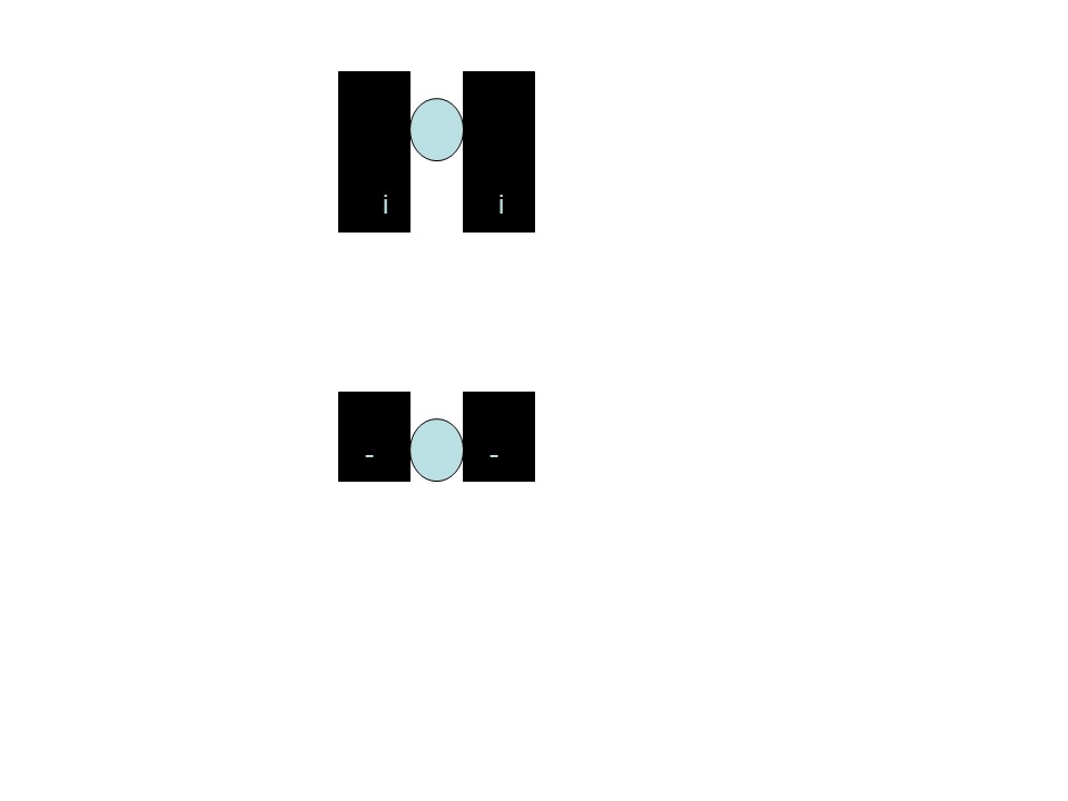
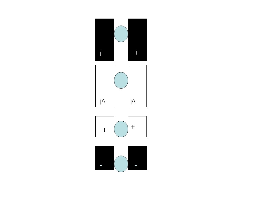
Now lets try some examples. What stages are the following cells?. The black chromosomes come from the father and the white from the mother. Ignore the symbols, they are too small to read and are not needed for this exercise.
The top one is?
The middle one is?
The bottom one is?
The top one is Meiosis 2. There are no maternal chromosomes at all. Therefore it can not be Mitosis. If it were M1, you would also see equal numbers of paternal and maternal chromosomes and you would see 4 sisters across.
The middle one is Meiosis 1. There are equal numbers of maternal and paternal chromosomes and there are 4 sisters across.
The bottom one is Mitosis. There are equal numbers of maternal and paternal chromosomes and there are not 4 sisters across,
When you go to the site, use the student login (this does not require any registration and put the code number where it says “enter Teacher’s room code”
It will ask for a name. You do not need to give your real name. When you are done with the exercise, it will say “teacher waiting or something similar. There will be no more activities at this site.
Be able to compare mitosis and meiosis in terms of processes and end results
The purpose of mitosis is to create cells that are genetically identical to the parent cells. Mitosis gives you cells that have 2 copies of each chromosome, just like the parent cells
The purpose of meiosis is to create cells that have 1 (not 2) copies of each chromosome so that after fertilization, the embryo will have 2 copies of each chromosome.
In both process, there is a preceding interphase which includes chromosome duplication.
Both processes have prophase, metaphase Anaphase and Telophase using the same cellular structures such as spindles and centrioles. Meiosis has 2 sets of these stages.
In order to assure that egg and sperm only get one of each chromosome, the homologues (members of a pair) line up during M1 and separate. During M1, crossing over occurs between homologues, this results in increased genetic variation
Crossing over does not occur in mitosis.
By Anchor207 (Own work) [CC BY-SA 3.0 (http://creativecommons.org/licenses/by-sa/3.0)], via Wikimedia Commons
Be able to distinguish homologues and sisters and know when in meiosis they separate
As said before Homologues have the same genes but may have different versions (Alleles) Homologues separate at M1. Sisters are initially (before recombination) genetically identical. They separate at M2. (They also separate during mitosis)
Be able to order the steps of meiosis
Chromosome Duplication (Before meiosis) Then Pairing of Chromosomes (Prophase of M1) Crossing Over (Prophase of M1) separation of Homologues (Anaphase of M1) separation of sisters (Anaphase of M2)
Know how meiosis leads to genetic variation (Know how recombination and independent assortment can increase the variation in gametes)
Recombination can lead to different combinations of alleles (gene variants) on the same chromosome
Independent assortment See page 263. Figure 13.11. See that there are 4 possible combinations of chromosomes when there are 2 pairs. As a rule, the number of possible combinations is 2^N (2 to the Nth) power where N is the number of pairs of chromosomes. Thus a male can have 2^23 possible combinations or over 8 million possible sperm. And that is without recombination!
Final exercise. //scratch.mit.edu/projects/embed/11743273/?autostart=false


 Note: Problems like the first one may appear in exams and quizzes. Problems like the second one will appear in Mastering Biology and the Group Worksheet.

 Just as in the evolutionary tree, in this diagram bonds that are grouped have features in common with bonds that are not grouped. For example The covalent bonds share electrons, while the ionic and hydrogen do not. What do hydrogen and ionic bonds have in common that is not in common with the covalent bonds?