Chapter 4 Objectives Fall 2020


Be able to list and recognize the principles of the cell theory
This video is useful and fun.

History of the Cell Theory

Be able to give applications of GFP

. In order to examine moving structures in living cells scientists took advantage of a jelly fish which makes a protein which glows green in fluorescent light. This protein is called GFP.(Green fluorescence protein) Using molecular tricks, one can fuse the GFP protein to other proteins. Where ever that other protein is, there will be a green signal. This video shows an example:

GFP in c. elegans development

In this video, a protein associated with chromosomes is labeled green. First, you can see the nucleii from the 2 parents  fuse together. Then the embryo starts cell division and you can follow the nucleii as this happens. The organism is C. elegans which is a tiny worm.


Cell Parts. Here are some activities to help you remember cell parts and functions

Here is a review video

Tour of the cell

Cell Parts

(Click on the flag and then, when prompted, click on the red dot associated with the structure)

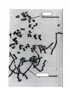
Two structures that easily get confused are the nucleus and the nucleolus


Nuclear Structure

Note the difference.

The function of the nucleolus is to help make ribosomes

The nucleus stores the chromosomes which have the genetic information for a cell. The nucleus is also needed for cell division and contains the nucleolus.

Know the pathway that membrane and secreted proteins take to get to or through the cell membrane. Know what happens at each step.

Exocytosis


Know the functions of cytoskeletal proteins including microtubules microfilaments and intermediate filaments.  Be able to give examples of how these elements contribute to movement of cells and of structures within cells and how they are involved in structure and shape of cells.


here is a video on cilia function

Cilia Function


Be able to give examples of how human disorders relate to defects in specific cell structures and/or proteins.

Here is a video on lysosomal storage disease

Lysosomal Storage Disease

EBS

This boy has a condition called EBS (Epidermolysis Bullosa Simplex), which causes severe blistering. This is a genetic condition which can be caused by defects in a number of proteins including keratin (an intermediate filament protein in the skin) and in components of the anchoring junctions. Based on what you have read about the functions of these components, why might a defect in them results in the blistering condition?


https://commons.wikimedia.org/w/index.php?curid=9793806

These are red blood cells  from someone who has a defect called Hereditary Spherocytosis. Normal red blood cells are disc shaped. In the microscope, they will usually shown a clearer area in the center. Abnormal red blood cells are rounder and do not have the clear area in the center.  Depending on the severity of the disease, the patient will have different numbers of normal and rounder red blood cells. The condition is genetic and is due to defects in a number of proteins which are known to associate with actin. Based on what you know about actin, explain the effect of the defect on the cells.


Extracellular Matrix; In addition to what is in the book, you may find this video quite informative

. Extra Cellular Matrix


Here is a useful review activity

Review Activity

Use the “match’ Function to do a speed test.


Cell Cycle Regulation And Cancer


Objective: Be able to predict what happens when cells from different phases are fused together.  Explain the results of fusion of interphase and M cells with respect to cyclin and MPF

Start by looking at this image

In this experiment cells from G1 phase are fused with cells from M phase

The cells from M phase (at top) have gone through S, therefore they have sisters.

The cells from G1 phase have not gone through S, do they have not replicated.

They have just started to condense. You can see them (not typical of G1 chromosomes) but they are not as condensed as the ones above.

Objective distinguish the roles of cyclin and CDKs in promoting the cell cycle 

Objective be able to interpret the cell fusion events in terms the functions of cylcin and CDK

Based on these results, researchers hypothesized the presence of a factor that induced M phase.  Years of genetic and biochemical research showed that this something is a factor called cyclin. Cyclin is a protein that accumulates at particular cell cycle stages and then are destroyed.  Different cyclins trigger different  stages.

Cyclin B is responsible for mitosis

The key here is that cyclin by itself does not cause M phase. Instead, it activates an enzyme called CDK. CDK stands for cell division Kinase. A kinase is an enzyme that adds phosphate. When CDK adds phosphates to chromosomes, they condense. This enzyme is also responsible for other prophase events like formation of a spindle and breakdown of the nuclear membrane.

For cell division, recall that there is a checkpoint a the G1/S boundary. In order for cells to advance to S, they need proteins called growth factors. For example, if you injure yourself platelets (a type of blood cell most known for clotting) releases a protein called Platelet Derived Growth Factor. This protein stimulates cells to divide to replace the dead ones.

Here is a video about hair growth factors (sorry, it is an ad, but it gets the point across)

Nanogen Hair Growth

Reception In cell division, the growth factor binds to a receptor. Two common types are Enzyme linked and G-protein linked. Both play roles in cell division control.

Here is an example showing an enzyme linked receptor from Open Stax

ART CONNECTION
This illustration shows two receptor tyrosine kinase monomers embedded in the plasma membrane. Upon binding of a signaling molecule to the extracellular domain, the receptors dimerize. Tyrosine residues on the intracellular surface are then phosphorylated, triggering a cellular response.
A receptor tyrosine kinase is an enzyme-linked receptor with a single transmembrane region, and extracellular and intracellular domains. Binding of a signaling molecule to the extracellular domain causes the receptor to dimerize. Tyrosine residues on the intracellular domain are then autophosphorylated, triggering a downstream cellular response.The transduction event starts when the enzyme is made active by addition of phosphate which happens when the receptor binds its ligand.Transduction: The next step with this enzyme is that it will add phosphates to other proteins and these will add phosphates to more proteins. The end result is amplification of a signal. It is sort of like our current epidemic. One person gets another person sick and that person gets 3 more people sick and soon there are thousands of sick people.In this system, the “people” are kinases, enzymes which add phosphates to other enzymes.
Response: These kinases activate enzymes and other proteins and lead to various results. One example is that some proteins go into the nucleus and tell cells to to make proteins like cyclins that trigger cell cycle events. A second example is a protein called Rb actively prevents cell division. Add a phosphate and the “brake” is released.This is shown in the diagram from Open Stax

ART CONNECTION
This illustration shows the regulation of the cell cycle by the Rb protein. Unphosphorylated Rb binds the transcription factor E2F. E2F cannot bind the DNA, and transcription is blocked. Cell growth triggers the phosphorylation of Rb. Phosphorylated Rb releases E2F, which binds the DNA and turns on gene expression, thus advancing the cell cycle.
(1) When no growth factors are present Rb sticks to a protein called E2F. This prevents E2f  from telling the cell to make proteins needed to perform S phase
(2) When growth factors are present, a series of phosphorylation reactions occur, that end up adding a phosphate to Rb (Right) This releases E2F and the cell makes S phase cell cycle proteins.
Here is a review question relevant to Rb. Click and answer
Kinase Question
Now imagine that there is no Rb protein present at all. Click on the question below and answer it.
Rb Question

So that will help us transition into the last part of this lesson:

Cancer.

Rb stands for Retinoblastoma, a protein that was originally identified as being involved in in a rare childhood eye cancer.

This is the cancer connection:

Most people have 2 copies of the gene that makes this protein, one from each parent. During one’s lifespan, however, it is possible that both of these copies are damaged. If that happens, then Rb can not stop cells from going to S when they shouldn’t and excessive cell division occurs: Cancer. Note that while this gene was originally shown to be important in eye cancer, it is involved in many other types as well.

Some people have 1 only one functional gene, because they inherited a mutant from one of their parents. Their cells are OK with one copy, but it only takes one error to mutate the “good” copy. As a result, they have a very high rate of cancers.

Genes like Rb are called Tumor Suppressors. They have the following characteristics

(1) The normal function of the protein is to prevent the cell from dividing when it shouldn’t.

(2) When both copies of the gene are mutated to a form that does not function  , cancer can result.

Here is another example from Open Stax, called P53. 50% of cancers have mutations in p53.

Mutated p53 genes have been identified in more than one-half of all human tumor cells. This discovery is not surprising in light of the multiple roles that the p53 protein plays at the G1 checkpoint. A cell with a faulty p53 may fail to detect errors present in the genomic DNA (Figure). Even if a partially functional p53 does identify the mutations, it may no longer be able to signal the necessary DNA repair enzymes. Either way, damaged DNA will remain uncorrected. At this point, a functional p53 will deem the cell unsalvageable and trigger programmed cell death (apoptosis). The damaged version of p53 found in cancer cells, however, cannot trigger apoptosis.

ART CONNECTION
Part a: This illustration shows cell cycle regulation by normal p53, which arrests the cell cycle in response to DNA damage, cell cycle abnormalities, or hypoxia. Once the damage is repaired, the cell cycle restarts. If the damage cannot be repaired, apoptosis (programmed cell death) occurs. Part b: Mutated p53 does not arrest the cell cycle in response to cellular damage. As a result, the cell cycle continues, and the cell may become cancerous.
The role of normal p53 is to monitor DNA and the supply of oxygen (hypoxia is a condition of reduced oxygen supply). If damage is detected, p53 triggers repair mechanisms. If repairs are unsuccessful, p53 signals apoptosis. A cell with an abnormal p53 protein cannot repair damaged DNA and thus cannot signal apoptosis. (Cell death) Cells with abnormal p53 can become cancerous. (credit: modification of work by Thierry Soussi).
A normal cell will respond to DNA damage by fixing the damage, and if the damage is too severe, the cell will die by apoptosis. (Kill the cell, spare the organism)A cell lacking the P53 gene will not repair damaged DNA and it will allow abnormal cells to keep dividing. This can lead to cancer.


Now we will talk about the second class of genes involved in cancer, activated oncogenes. An example is a gene called HER2 which stands for Human epidermal growth factor receptor. Below shows what happens in a normal person.This illustration shows two receptor tyrosine kinase monomers embedded in the plasma membrane. Upon binding of a signaling molecule to the extracellular domain, the receptors dimerize. Tyrosine residues on the intracellular surface are then phosphorylated, triggering a cellular response.In some cancers (notably some forms of breast cancer), the receptor (because of a mutation) no longer needs the growth factor (The signalling molecule in red) to bind to the receptor to be active. This sets off the chain of events that leads to uncontrolled cell division.This cases illustrates some general principles of activated oncogenes (compare to tumor suppressors)
Here is a video discussing HER2
HER 2
(1)The normal function of the gene is to promote cell division(2) Mutated versions are not properly regulated or too much is made
(2) Only 1 copy of the gene has to be mutated(3) They are rarely, if ever, inherited from parents.

Now answer this question (click on the link)

BRCA1 question

Here is some good news about cancer therapy.

Emily was one of the first to have this therapy, but now it is more widely. Former President Jimmy Carter, who is over 90 was successfully treated in this manner:

Cancer Immunotherapy.


The Cell cycle lesson


Overview Video for eukaryotic cell cycle: This gives some basics on the overall process of cell reproduction and is visually striking

Cell Division Video
Objective: Know what happens in each stage of the cell cycle including each stage of mitosis Be Able to recognize cells at each stage. 

Match the images to the stages!

This activity was made by a former student Athraa Kamil as a “Miss One, Make One” activity

Cell Cycle Matching

Here is a video on mitosis with embedded questions

Mitosis Video with questions


Objective: Be able to determine the mitotic index of cells

Objective: Be able to determine cell cycle stage lengths from data about stage distribution

Example. The total length of the cell cycle for a onion root tip cell is 30 hours.  You find 120 cells in interphase, 30 in prophase, 15 in metaphase  25, in anaphase, and 15 in telophase or cytokinesis. What is the mitotic index? What is the length of prophase?

The mitotic index is the percent of total cells in mitosis. In this case, there are 200 total cells and 80 are in the stages of mitosis, so  the mitotic index is 80/200=.4

To determine the length of a particular stage first determine the fraction of cells in that stage. In this case 30/200 or  15% of the cells are in prophase. We assume that the proportion of cells in a particular stage is directly related to the length of that stage (the more cells at a particular stage, the longer that stage must be. If the total cell cycle is 20 hours and 15% are in prophase, then 15% X 30=4.5 hours.

Objective: Be able to order the steps of the cell cycle and mitosis


Be able to label chromosomes including sister chromatids, centromeres and the kinetechores


Objective Be able to distinguish sister chromatids from homologues


For a given chromosome, every individual has one homologue from each of their parents  A and a are different alleles (different versions of the same gene; brown and blue eyes for example)

Sisters and Homologs in Mitosis

Objective: Know the role of the following in the cell cycle: Centromere, Kinetochore, Centriole, Centrosome, Spindle, Contractile Ring

Note that centromere, centrosome and centriole all sound alike and are easy to confuse. Only the centromere is directly associated with chromosomes.


The centromere is the   structure at which 2 sisters are attached. It is also attached to the Kinetochore which links the spindle to the chromosome.

Here is a useful slideshow on chromosome structure

Chromosome structure review

Centrioles are barrel shaped structures from which microtubules grow.  The centrosome contains the centrioles and some other components which help form microtubules


Plants do not typically contain centrioles, but they do contain centrosomes with  This appears sufficient to form spindles


Chapter 8


For each of the following, know where they are made and where they are used  (The light cycle or Calvin Cycle): ATP, NADPH, NADP+, Oxygen, Carbon Dioxide, Sugars,  Part of the table is filled in for you. Can you fill in the rest?  

Compound Light Cycle Calvin Cycle
ATP Made Used
NADPH
NADP+
Oxygen Made Not made or used
Carbon Dioxide
Sugars


The link below is an exercise to put steps of photosynthesis in order

Photosynthesis Order


Be able to interpret an action spectrum and distinguish it from an absorbance spectrum

Plants move toward light. The image below shows how the relationship between the wavelength of light and the degree of movement. This is an action spectrum, because it measures a plant response as a function of wavelength. This can be compared to an absorbance spectrum which measures absorbance of light as a function of wavelength (like our first lab). What would you predict about the absorbance spectrum of a pigment that was responsible for the response in the graph below? What would be a good title for this graph?

Action Spectrum for phototropism


Be able to label a chloroplast and show where the Calvin cycles and light cycles take place.


Be able to compare respiration with photosynthesis. Which processes and components do they have in common?


Chapter 6 Objectives

To understand the basic energy rules, view the slideshow

Energy Rules

Here is a video on thermodynamics and perpetual motion machines

Perpetual Motion machines and Thermodynamics

Enzymes:  First watch this video

Enzyme Video

Know what substrates are and how they relate to enzyme catalyzed reactions.

Example what is the substrate in this enzyme catalyzed reaction

Lactose→Glucose + Galactose?

The enzyme in this reaction is lactase

Know how enzymes catalyze biological reactions, specifically their effects on activation energy. 

Another concept in how enzymes speed up reactions is activation energy. Imagine you have a homework assignment due next week. You could do it now, or you could put it off until right before it is due.  The activation energy is the energy needed to get the process started. Note that the homework might take you an hour whether you do it now or later. The activation  energy is just what it takes to get started. Enzymes decrease activation energy as shown in the diagram in the link

Activation Energy


Note that that the ΔG (The overall energy) does not change whether or not the enzyme is present. Enzymes also have no effect on the equilibrium of a reaction; adding an enzyme does not shift the reaction to the left or right.  It does change how quickly the reaction reaches equilibrium.

Understand how compounds that bind to the active site or allosteric site can alter enzyme function

Many drugs can inhibit enzyme function by binding to the active site. Here is an example

HIV Drug Action

The key point here is that the inhibitor binds very strongly to the active site preventing the substrate from binding. Because both the inhibitor and substrate bind to the same site, the binding is said to be competitive.  If the concentration of substrate increases, the inhibitor is less effective.

We have already mentioned active sites, they bind to the substrate(s) of a reaction. Some enzymes also have allosteric sites. These do not bind to substrates but they bind to compounds that can turn on (activators) or turn off (inhibitors) an enzyme as shown in this video

Allosteric regulation of enzymes

Non-competitive inhibitors bind to the allosteric site.  Adding more substrate has no effect on allosteric inhibitors since the inhibitor changes the active site to be “closed”.

Know what feedback inhibition is and how it can be used to control amino acid synthesis.

Feedback inhibition

Here is an example of how this works to control amino acid levels

Feedback inhibition o f amino acid synthesis


Introduce Yourself!

Give your name, where you are from and what most interests you about Biology. Also say whether what has helped you in online learning (even if it was just the end of last semester or a bit in high school).

Put your answer as a reply.

I have to approve all posts, so you will not see your reply immediately.

Chapter 3 Objectives Summer 2020


Here are some exercises to help you understand the week 2 objectives

Be able to identify carbons and hydrogens in organic compounds

Watch this video

Be able to determine whether 2 structures are isomers and whether they are structural, geometric or enantiomeric isomers.

For 2 structures to be isomers, they have to have the same molecular formula

Watch the video. Note that geometric isomers are a type of diastereomers. Geometric isomers are distinguished by having a double bond and a different arrangement of atoms around he double bondd


Be able to identify functional groups.

Click on the green flag to start. The faster you do this, the higher your score
//scratch.mit.edu/projects/embed/112453626/?autostart=false

Be able to recognize and give functions for the following biomolecules: Monosaccharides, disaccharides, polysaccharides. Fats, phospholipids, steroids. Amino acids, proteins.

The following exercise will help you recognize the key differences between carbohydrates and lipids.Click on the green flag and observe which structures are lipids and which are carbohydrates.
Click next and then click the appropriate button
Continue hitting next and choosing the correct button

Carb and Lipid Test

Here is a second activity for carbs and lipids

Carbs and lipids

Be able to distinguish primary, secondary, tertiary and quaternary structure

This can be difficult for people to remember.  A few key points

  1. The levels represent interactions within a protein. They do not necessarily represent steps in protein folding .
  2. You should know (1) What types of bonds are used and (2) What parts of the amino acids are involved. Be sure you can identify R groups and the backbone portions of amino acids.

Here is a protein structure review

Protein Activities

5. Watch the following video and answer the embedded questions:
https://edpuzzle.com/embed/media/575adfdf98ce82292d1b1a4d


Here is a review of macromolecules you need to know

https://edpuzzle.com/embed/media/54e792d9073086a942ceed2f


css.php